
The microscopic world is full of wonders and details that are too small for the naked eye to see. With the help of a microscope, however, we can dive into this world and discover the hidden beauty of nature. But how do you observe through a microscope? In this article, we’ll explore the basics of using a microscope and share tips on how to get the most out of your observations. Whether you’re a student or a curious learner, this guide will help you unlock the secrets of the microscopic world. So let’s dive in and discover how to observe through a microscope!
Contents
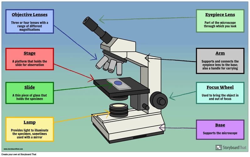
What is a Microscope?

A microscope is an instrument that is used for observing things that are too small to be seen with the naked eye. It uses a combination of lenses to magnify an object or a sample, allowing us to view it in detail and study its features.
There are different types of microscopes available, such as compound microscopes, stereo microscopes, and electron microscopes. Compound microscopes are commonly used in schools, laboratories, and research centers for magnifying specimens like cells, bacteria, and tissues. Stereo microscopes, on the other hand, are used for studying larger objects like minerals, insects, and flowers. Electron microscopes, which use a beam of electrons to create an image are used for studying very small objects like atoms and viruses.
The magnification power of a microscope can range from a few times to thousands of times the actual size of the object. A higher magnification power often results in a decrease in the field of view or the area that can be viewed through the microscope. Microscopes are also equipped with different features like focus knobs, eyepieces, and illumination sources to ensure that the object is in focus and clearly visible.
In summary, a microscope is a tool used for observing things under a microscope, revealing details that cannot be seen with the naked eye. It is important for different fields including science, medicine, and research, allowing us to see and study things at the microscopic level.
Preparing to Observe Through a Microscope

Choosing a Microscope
Choosing the right microscope is essential for proper observation. There are different types of microscopes, such as compound microscopes, stereo microscopes, digital microscopes, and more. The type of microscope you choose will depend on what you want to observe and how you want to observe it. Don’t just choose any microscope; you must select a microscope that can deliver accurate results.
When selecting a microscope, consider the magnification, resolution, and the quality of the lenses. Higher magnification provides a clearer view of the object under observation, while good lenses and resolution offer a better and sharper view.
Preparing the Sample
Preparing your sample correctly is just as crucial as choosing the right microscope. If your sample is not well-prepared, you will not get an accurate result, and the microscope will not reveal its wonders.
First, ensure that your sample is clean, dry, and flat. Dust, moisture, and wrinkles can obstruct the view of the object under observation. Then, put the object on a slide and use a coverslip to protect the object and the lens. Be careful when handling your samples to avoid damage, as even a small scratch can affect the clarity of your image. Additionally, if you plan on observing living organisms, you must ensure that the sample stays alive and does not dry out during the observation.
In summary, preparing to observe through a microscope involves choosing the right microscope and preparing the sample correctly. Both are equally important and require attention to detail for accurate results.
How to Use a Microscope

Adjusting the Focus
Before beginning your experiment, it’s important to ensure that your microscope is focused properly. To adjust the focus, locate the coarse focus knob and turn it until the objective lens is approximately 1 inch away from the stage. Then, adjust the fine focus knob to bring the sample into clear view. Repeat this process as needed during your experiment to keep the sample in focus.
Magnification
One of the most important considerations when using a microscope is the level of magnification you need. High magnification is great for observing small details, but may result in a reduced field of view. To adjust the magnification level, locate the nosepiece and rotate it until the desired objective lens is in place. Then, adjust the focus as needed.
Illumination
Good illumination is crucial for clear observations when using a microscope. To adjust the illumination, locate the light source and adjust the intensity level. Some microscopes feature an adjustor that allows you to move the light source closer or farther away from the sample to get the desired level of illumination.
Illumination Type
There are several illumination types commonly used for microscopes: bright-field, dark-field, and phase contrast. Bright-field is the most common and uses uniform illumination, while dark-field uses angled illumination to create contrast. Phase contrast uses differences in refractive indexes to create contrast, making it ideal for observing transparent samples.
Positioning the Sample
Properly positioning your sample is key to getting clear observations with a microscope. Make sure the sample is firmly and evenly mounted on the slide, and use the stage controls to move the slide until the desired area is in the field of view. If you need to move the slide while observing, be sure to keep track of the sample’s position and use the stage controls to find it again.
Observing Through a Microscope
Viewing Living Organisms
When observing living organisms through a microscope, the first thing you need to do is to prepare a slide using a wet mount technique, which involves putting a drop of water on the slide and adding a specimen. Once the slide is prepared, place it on the microscope stage and adjust the focus using the coarse and fine focus knobs. To examine the specimen further, you may need to adjust the magnification or change to a higher power objective lens. It’s essential to avoid using too much light because it can damage or kill the specimen.
Viewing Inorganic Samples
Viewing inorganic samples with a microscope requires a different technique. To prepare a slide with an inorganic sample, use a dry mount technique. This involves placing the sample on a slide and fixing it in place with a coverslip. When observing inorganic samples, it’s essential to use reflected light instead of transmitted light. Reflected light provides a clearer image of the sample’s surface. Like with the living organisms, adjust the focus and magnification to get a better view of the sample.
Photomicrography
If you want to capture images of what you observe under a microscope, photomicrography is the way to go. Photomicrography is the process of taking pictures through a microscope. You can achieve this by attaching a camera to the microscope or using a specialized photomicrography microscope. It’s essential to use a high-quality camera and a tripod to avoid blurry images. Additionally, use appropriate lighting to enhance the clarity of the image.
Cleaning and Maintaining Your Microscope

- Don’t touch the lenses: The most important step in maintaining your microscope is to avoid touching the lenses. Even fingerprints can damage the lenses and cause blurry images.
- Clean the lenses: To remove dust and dirt, use a soft-bristled brush to gently remove any debris. If the debris is stubborn, use a lens cleaner or alcohol-based solution to gently clean the lenses.
- Check the bulbs: Microscopes rely on a light source to illuminate the specimens being viewed. Check the bulbs regularly to make sure they are working properly, and replace them as needed.
- Use protective covers: When not in use, protect your microscope by using dust covers or carrying cases. This will prevent dust and debris from accumulating on the lenses and other parts of the microscope.
- Store properly: Proper storage is key to maintaining the longevity of your microscope. Store it in a dry, cool place away from direct sunlight to avoid any damage to the lenses and other parts.
- Regular maintenance: Regularly inspect your microscope for any scratches or cracks on its lenses, wear and tear on other parts, and proper alignment of the lenses. Make any necessary repairs or adjustments immediately to prevent any further damage.
In summary, taking proper care of your microscope will ensure you get the best possible results and extend the longevity of your instrument. By following these simple guidelines, you can enjoy the beauty and wonder of the microscopic world for years to come.
Different Types of Microscopes

Microscopes are essential scientific equipment used for observing small objects, cells, microorganisms, and other microscopic specimens. There are different types of microscopes, each designed for specific uses. Here are some of the commonly used types of microscopes:
- Compound microscope: This type of microscope is used for observing thin specimens such as cells, microorganisms, and tissues. It has two or more lenses, which magnify the specimen by bending the light that passes through it.
- Stereomicroscope: Also known as a dissecting microscope, this type of microscope is used for studying larger specimens such as insects, rocks, or plants. It has two lenses, which provide a 3D view of the specimen, allowing the observer to see its surface and internal structures.
- Electron microscope: This type of microscope uses a beam of electrons to produce images of specimens. It provides a higher resolution and greater magnification than a compound microscope, making it suitable for studying extremely small objects such as viruses, bacteria, and molecules.
- Scanning probe microscope: This type of microscope uses a probe to scan the surface of a specimen and create images with atomic-scale resolution. It is used for studying materials science, nanotechnology, and other fields where high precision is essential.
- Confocal microscope: This type of microscope is used for producing 3D images of specimens, typically in biology and medicine. It uses a laser to scan the specimen and create a series of images, which are then combined to create a 3D view.
In conclusion, microscopes are an essential tool for scientific research, and the type of microscope used depends on the specimen being studied and the purpose of the study. By understanding the different types of microscopes, researchers can choose the most appropriate tool for their research, leading to breakthroughs and discoveries in various fields.
Potential Hazards
Observing through a microscope may seem harmless, but there are several potential hazards that you should be aware of to ensure your safety.
| Hazard | Description | Precautionary Measures |
|---|---|---|
| Eye Injury | High-intensity light from the microscope can cause eye damage, which can lead to temporary or permanent vision loss. |
|
| Electrical Shock | If the microscope is not properly grounded, there is a risk of electrical shock. |
|
| Chemical Exposure | Some staining agents used in microscopy can be toxic if inhaled or ingested. |
|
| Physical Injury | Microscope slides and coverslips are fragile and can break, causing cuts or punctures. |
|
By being aware of these potential hazards and taking necessary precautions, you can safely explore the microscopic world and marvel at its wonders.
Frequently Asked Questions
What type of microscope should I use for observing the microscopic world?
To observe the microscopic world, using the right type of microscope is crucial. Here are some common types of microscopes that you can use:
- Compound Microscope: This type of microscope is suitable for observing small and thin specimens, including cells, tissues, and bacteria. It has high magnification and resolution, making it perfect for scientific research and laboratory work.
- Stereo Microscope: Also known as a dissecting microscope, this type of microscope is ideal for observing larger and thicker specimens, such as insects, rocks, and plants. It has a lower magnification and a 3D view, making it easier to observe the details of the specimen.
- Electron Microscope: This type of microscope is used for observing ultra-small objects, including molecules, viruses, and nanostructures. It uses a beam of electrons to magnify the specimen, providing a higher level of detail than other types of microscopes.
It is important to choose the right microscope for your intended observation. While compound and stereo microscopes can be used for educational purposes, electron microscopes are mainly used for advanced scientific research. Consider the magnification, resolution, and features of each type of microscope before making a purchase or selecting one for your experiment.
What are the different magnification levels available for a microscope?
Microscopes come in different types and sizes, and each type has its own set of magnification levels. Here are some of the commonly available magnification levels for microscopes:
- Low power magnification: This level of magnification is used to observe larger specimens, such as insects or plant cells, and range from 40x to 100x.
- Medium power magnification: This magnification level is used to observe smaller specimens, like blood cells or small organisms, and typically ranges from 100x to 400x.
- High power magnification: This magnification level is used to observe the fine details of a specimen, such as individual cells or bacteria, and ranges from 400x to 1000x.
- Oil immersion magnification: This is the highest level of magnification, and is achieved by placing a drop of oil on the slide and using a lens with a magnification of 1000x or more. This technique is used for observing very small specimens, such as viruses.
The magnification level required depends on the size and detail of the specimen being observed. It is important to adjust the focus and lighting settings accordingly to ensure a clear and accurate observation. With the right magnification level and technique, the microscopic world can reveal a vast and fascinating universe that is otherwise invisible to the naked eye.
What materials are best for preparing samples for viewing under a microscope?
When it comes to preparing samples for viewing under a microscope, choosing the right materials is crucial. Specimens need to be properly prepared to obtain clear, sharp images. Here are some of the best materials for preparing samples for viewing under a microscope:
- Microscope slides are essential for holding specimens in place. They are typically made of glass and come in a variety of sizes and thicknesses.
- Coverslips are thin pieces of glass that are placed over specimens to protect them and keep them in place. They come in different sizes and thicknesses and can be used with a drop of water or mounting medium.
- Mounting medium is a substance used to permanently preserve a specimen on a microscope slide. It can be made of various materials depending on the type of specimen being viewed.
- Stains can be used to increase contrast and highlight specific structures in a specimen. There are a variety of stains available for different types of specimens.
Using the right materials for preparing samples can greatly enhance the clarity and detail of the images obtained under a microscope.
What types of lighting should I use to best observe the microscopic world?
- Brightfield Illumination: This is the standard lighting type used in most microscopes. It illuminates the specimen with a bright, white light from below. Brightfield illumination is best for observing specimens that are stained or have a high contrast.
- Darkfield Illumination: This type of lighting is used when you want to observe small, transparent specimens that would be difficult to see under brightfield illumination. It works by illuminating the specimen with a cone of light from the side, which creates a contrasting, dark image.
- Phase Contrast Illumination: This type of lighting enhances the contrast of transparent specimens by amplifying the differences in refractive index between different parts of the specimen. It works by splitting the light into two beams, one of which goes through the specimen and the other around it. This creates an image with dark and light areas that reveal the internal structure of the specimen.
- Fluorescence Illumination: This type of lighting is used with specimens that have fluorescent properties. It works by illuminating the specimen with a specific wavelength of light, which causes the specimen to emit a different colored light. Fluorescence illumination is commonly used in biological research to study cellular and molecular processes.
In order to get the best observation results, it is important to choose the appropriate lighting for the specimen you are observing. By understanding the different types of lighting available, you can optimize your microscope’s lighting to achieve the best results possible.
What Safety Precautions Should I Take When Using a Microscope?
- Always wear protective equipment: Safety glasses or goggles should always be worn when using a microscope to prevent any particles or samples from getting into your eyes.
- Handle the microscope with care: Always carry the microscope with both hands, and be gentle when adjusting the knobs and lenses to prevent any damage to the microscope. Avoid touching the lenses with your fingers, and use lens paper to clean them instead.
- Be cautious with the light source: Avoid looking directly into the light source of the microscope, which could cause eye damage. Also, turn off the illumination when you are finished using the microscope to prevent excess heat or damage to the light bulb.
- Dispose of hazardous materials: Dispose of any hazardous materials appropriately, following the proper protocol for disposal. Never dispose of any chemicals or samples down the sink or drain.
- Always work in a well-ventilated area: Many samples and chemicals used in microscopy can emit fumes that can be harmful if inhaled. Always work in a well-ventilated area or use a fume hood when necessary.
By following these safety precautions, you can help ensure a safe and enjoyable experience when using a microscope.
Conclusion
Exploring the microscopic world can be a fascinating experience. With the right equipment, a bit of practice, and patience, you can learn to observe and appreciate the many wonders that exist on a tiny scale. Microscopy is an exciting way to expand your understanding of the natural world and uncover mysteries that may have gone unnoticed.


