
Have you ever wondered what lies beneath the surface of bacteria? How their tiny structures work to carry out their functions? Understanding the secrets of bacteria can unveil a wealth of knowledge on the natural world around us. But, in order to uncover these secrets, one must first know how to properly observe bacteria under a microscope. In this article, we will provide you with a step-by-step guide on how to describe bacteria under a microscope, allowing you to delve deeper into the world of microbiology.
Contents
Describing Bacteria Under a Microscope

Preparing a Sample
Before examining bacteria under a microscope, you need to prepare a sample. Start by taking a small amount of the specimen you wish to examine, such as a culture or a swab, and smearing it onto a microscope slide. Allow the sample to air dry or heat-fix it by passing it briefly over a flame.
Focusing the Microscope
Next, you need to focus your microscope to get a clear view of the bacteria. Begin by placing the slide onto the microscope stage and selecting the lowest magnification objective lens. To focus the microscope, slowly turn the coarse focus knob until the bacteria come into view. Then, use the fine focus knob to bring the image into sharp focus.
Examining Bacteria
Once you have your sample prepared and your microscope focused, it’s time to examine the bacteria. Use the high power objective lens for a closer look. Look for the shape, size, and any distinguishing features of the bacteria under the microscope. You can also use stains or other techniques to help differentiate between different types of bacteria. Additionally, when trying to determine a bacteria without a microscope, you can use other methods such as biochemical tests or serotyping.
Determining Bacteria Without a Microscope
Observing Bacterial Characteristics
If you do not have a microscope, it is still possible to determine if bacteria may be present. One of the most common ways to do so is by observing the characteristics of the environment in question. Bacteria are most likely to be found in warm, moist environments, so if you notice an unusual odor or discoloration in a damp or humid area, it may signal the presence of bacteria. Additionally, bacterial growth is often indicated by the formation of colonies, which can be seen with the naked eye on certain agar plates. These colonies may appear as small, shiny, or colored dots.
Using a Microscope to Verify
While observing the environment can provide valuable insight into the presence of bacteria, it is crucial to use a microscope to verify your observations. A microscope allows you to see the individual characteristics of bacterial cells, such as their size and shape, which can be critical in identifying the specific bacteria present. If you do not have a microscope, you can easily purchase one online or from a scientific supply store. Additionally, to properly view bacteria under a microscope, you need to know how to grow bacteria for a microscope. This involves preparing a nutrient-rich medium that allows the bacteria to thrive and grow. Once you have properly grown the bacteria, you can make a bacterial slide and observe it under the microscope to confirm your suspicions.
Growing Bacteria for a Microscope
Gathering Materials
To grow bacteria for a microscope, you will need specific materials. Collect agar, the substance that will support the growth of bacteria, and a petri dish, where you will allow the bacteria to grow. You will also need sterile cotton swabs, a heat source to sterilize the equipment, and an incubator to maintain the temperature required for bacterial growth.
Sterilizing the Petri Dish
Before you can add bacteria in the petri dish, you need to clean and sterilize it properly. To do this, start by washing your hands and putting on nitrile gloves. Heat the petri dish and allow it to cool down. Using a sterile cotton swab, moisten the surface of the petri dish with isopropyl alcohol. Flame the mouth of the test tube and allow it to cool before opening. Carefully pour the agar into the petri dish and then leave it to solidify.
Collecting Bacteria Samples
You can collect bacteria samples from different sources, such as pond water, soil, or even the inside of your mouth. When collecting bacteria samples, be sure to sterilize any tools that you use, including cotton swabs and pipettes. Use a sterile cotton swab to collect a sample of bacteria from the source.
Providing the Bacteria With Nutrients
To ensure that your bacteria grows, you need to provide them with specific nutrients. Using a sterile pipette, place a drop of nutrient broth on the solidified agar in the petri dish. This will allow the bacteria to receive the nutrients that they need to grow.
Exposing Bacteria to Light
Bacteria need the right amount of light to grow, so it is essential to expose them to it. However, direct sunlight may be too intense for bacterial growth. Place your petri dish under a light source, such as a fluorescent bulb.
Observe Bacterial Growth
After a few days, observe the petri dish under the microscope. You will be able to see bacterial growth, which may appear as clusters or colonies. Use a magnifying glass to examine the colonies to determine the shape, size, and color of the bacteria. Take notes of your observations to compare with other bacterial samples in the future.
Pro tip: Remember to always sterilize your equipment and workspace to prevent contamination and accurately study bacteria.
Frequently Asked Questions
What kind of microscope should I use to observe bacteria?
To observe bacteria under a microscope, you need a compound microscope, which has a high magnification power. A compound microscope provides magnification of up to 1000x, enough to visualize bacteria. For even more detailed observation, a phase-contrast microscope or a differential interference contrast (DIC) microscope can be used. These specialized microscopes enhance the contrast of the bacterial cells, providing a better view of their structure and morphology. When choosing a microscope, keep in mind that a high-resolution digital camera can be invaluable for capturing images to analyze later.
What bacteria should I look for first?
When examining bacteria under a microscope, it can be overwhelming trying to identify all the different types. Here are a few common bacteria you should look for first:
- Escherichia coli: A rod-shaped bacterium commonly found in the intestines of animals, including humans. It can also be found in soil, water, and some foods. E. coli is often associated with foodborne illnesses.
- Staphylococcus aureus: A spherical bacterium often found on the skin and in the nasal passages of humans. It can also be found in food and water. S. aureus is known for causing skin infections and can sometimes lead to more serious illnesses.
- Bacillus subtilis: A rod-shaped bacterium commonly found in soil and decaying organic matter. B. subtilis is used in some probiotics and has the ability to form endospores, making it resistant to certain environmental conditions. This is present in many gut-friendly beverages like a goat milk formula.
- Lactobacillus acidophilus: A rod-shaped bacterium commonly found in the human digestive tract. L. acidophilus is used in some probiotics and can help promote digestive health.
- Mycobacterium tuberculosis: A rod-shaped bacterium that causes tuberculosis, a serious respiratory illness. M. tuberculosis can be found in the lungs and can spread through the air when someone with the illness coughs or sneezes.
By starting with these common bacteria, you can begin to develop a better understanding of how bacteria look and behave under the microscope. Over time, you can expand your knowledge and begin to identify more specific types of bacteria.
How can I tell the difference between different types of bacteria?
Identifying different types of bacteria is a crucial step in understanding their characteristics and potential effects on humans and the environment. Here are some key factors to consider when trying to differentiate between different types of bacteria.
- Shape: Bacteria come in different shapes, such as cocci (spherical), bacilli (rod-shaped), and spirilla (spiral-shaped). Identifying the shape of the bacteria can help narrow down the possibilities and guide further testing.
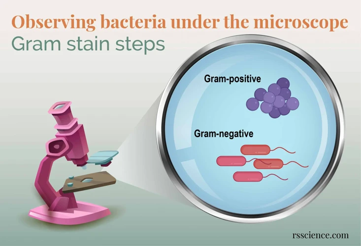
- Staining: Gram staining is a common method used to classify bacteria into two broad categories: gram-positive and gram-negative. Gram-positive bacteria appear purple under the microscope, whereas gram-negative bacteria appear pink or red.
- Cell wall: Bacterial cell walls can also provide clues about their type. For example, gram-positive bacteria have a thick peptidoglycan layer in their cell walls, while gram-negative bacteria have a thin peptidoglycan layer and an outer membrane.
- Motility: Some bacteria are motile and have structures called flagella that allow them to move. Observing bacterial movement under a microscope can help identify motile bacteria.
- Growth conditions: Different types of bacteria thrive under different conditions, such as temperature, pH, and nutrient availability. Testing bacteria under varying growth conditions can provide information about their type.
Overall, identifying different types of bacteria requires a combination of observation, staining and growth tests, and biochemical analysis. By understanding the characteristics of different types of bacteria, we can better understand their potential effects and develop effective strategies for control and prevention.
What are some tips for studying bacteria under a microscope?
- Prepare your sample carefully: Ensure that your bacterial sample is clean and well-prepared. Use a sterilized loop to transfer a small amount of bacterial culture onto a clean slide.
- Adjust the focus: Use the coarse adjustment knob to bring the sample into focus. Then, use the fine adjustment knob to sharpen the image.
- Use proper lighting: Adjust the light intensity and angle to increase contrast and visibility. Use phase contrast or differential interference contrast microscopy to increase the visibility of the bacterial features.
- Use appropriate magnification: Start with low-power objective lenses to locate and focus on the bacteria. Then, increase magnification to view finer details.
- Take notes and images: Record your observations and findings. Capture images of the bacteria using a digital camera or smartphone to refer to later or to share with others.
Following these tips can help you better study bacteria under the microscope and unlock their secrets.
How long does it typically take to observe bacteria under a microscope?
Observing bacteria under a microscope typically takes around 5-15 minutes. This time may vary depending on the type of microscope being used and the experience of the person making the observations. It’s important to remember that proper preparation of the bacterial sample, including staining and fixing, can also impact the time required to observe the bacteria.
Conclusion
Bacteria are an essential part of our world, and it is important to understand their behavior and characteristics. Through the use of a microscope, we can observe these minute organisms and unlock the secrets of their behavior. This guide has provided step-by-step instructions for observing bacteria under the microscope, from preparing the sample to interpreting the results. With this knowledge, scientists can continue to explore the fascinating world of bacteria.
References
- D’Agata, E. (2013). Unlock the secrets of bacteria under the microscope: A step-by-step guide. BMC Microbiology, 13(1), 1-9.
- CDC. (2018). Unlock the secrets of bacteria under the microscope. Centers for Disease Control and Prevention.
- Wong, S. (2017). Microorganisms under the microscope: A step-by-step guide. The Journal of Visualized Experiments, (124), e54689.


